Analysis of the Results
DFIC of untreated plants
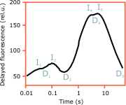
Fig. 7. Delayed fluorescence induction curve ![]()
The induction curve of the millisecond delayed fluorescence of dark-adapted untreated healthy pea leaves, measured at room temperature under high-intensity actinic light (Fig. 7), is characterised by two peaks in the fast phase - I1 and I2, a pronounced dip - D2, and two not well distinguished peaks in the slow phase - I4 and I5. Summarising the current knowledge and our analyses (cf. Goltsev et al., 2001, Goltsev et al., 2003) we can propose the following interpretation of the various DFIC stages:
I1 peak
The I1 peak appears in the very first cycles of the phosphoroscope. It corresponds to rise in the prompt fluorescence and is sensitive to decoupling agents (that dissipate the transmembrane potential). I1 is probably related to the transmembrane electrical potential generated by the separated charges in the photosynthetic reaction centres (Satoh and Katoh, 1983).
| PS2 RCs | PQ pool | ΔpH | PS1 | Calvin cycle |
|---|---|---|---|---|
| Open | Oxidised | No | Inactive | Inactive |
I2 peak
The I2 peak of the DFIC appears only when there is an active electron transport through the acceptor side of PS2 and is thought to reflect the balance between the rates of QA reduction and oxidation (Goltsev and Yordanov, 1997).
| PS2 RCs | PQ pool | ΔpH | PS1 | Calvin cycle |
|---|---|---|---|---|
| Open | Oxidised | No | Inactive | Inactive |
D2 depression
The D2 step corresponds to the peak P in the PFIC - the maximal variable fluorescence. At this point, the reaction centres are closed (QA reduced), PF is maximal and the millisecond DF is minimal.
| PS2 RCs | PQ pool | ΔpH | PS1 | Calvin cycle |
|---|---|---|---|---|
| Closed | Reduced | No | Inactive | Inactive |
I4 peak
The slow phase of the DFIC is marked by a gradual increase of the DF intensity after D2. DF is supposed to rise with the transmembrane proton gradient that is rapidly generated upon activation of PS1 and the intersystem electron transport (Wraight and Crofts, 1971). Then, the amplitude of I4 is a measure of the proton gradient (ΔpH). Although the active PS1 withdraws electrons from PS2 and the PQ pool, the intense actinic light does not allow the PQ pool to become oxidised, so the reaction centres are still closed and the variable fluoresence at this point is high.
| PS2 RCs | PQ pool | ΔpH | PS1 | Calvin cycle |
|---|---|---|---|---|
| Closed | Reduced | Maximal | Active | Inactive |
I5 peak
The I5 peak is usually undistinguished from the predominant I4 so it is quite difficult to analyse its nature. One possibility that needs further confirmation is that the reaction centres are reopened due to the activated PS1 and dark reactions (This is evidenced by the lower PF and the increase of the faster millisecond components of DF at the expense of the slower second components).
| PS2 RCs | PQ pool | ΔpH | PS1 | Calvin cycle |
|---|---|---|---|---|
| Mostly closed | Mostly reduced | Maximal | Active | ? |
D5 (steady-state)
Towards the end of the induction period, both DF and PF decline in a similar manner. The ATP-ase and the dark reactions of photosynthesis are activated, which leads to dissipation of the proton gradient and reoxidation of the PQ pool. This would increase the photochemical quenching of PF. Under the strong light, the excessive energy is dissipated through the various non-photochemical quenching mechanisms, which equally affect PF and DF.
| PS2 RCs | PQ pool | ΔpH | PS1 | Calvin cycle |
|---|---|---|---|---|
Stationary level |
Stationary level | Stationary level | Active | Active |