Delayed Fluorescence
Induction curves of delayed fluorescence
Like the variable fluorescence, DF undergoes a series of transient changes during the dark-to-light adaptation of the photosynthetic sample, which are called DF induction (Malkin, 1977). The induction curves of delayed fluorescence (DFIC) are reflect the electron transport capacity, the rate of photosynthesis, the level of the transmembrane proton gradient and the integrity of the thylakoid membrane, etc. On the other hand, the shape of the DFIC depends on the specific conditions of registration — the ambient temperature, the duration of the dark adaptation period, the intensity of the actinic light and the duration of the phosphoroscope cycles. Unfortunately these factors make the comparison of DFIC registered in different laboratories very difficult.
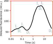
Fig. 7. Delayed fluorescence induction curve ![]()
A typical DFIC is characterised by several peaks (maxima) separated by depressions (minima). They are best visualised when the DFIC is plotted on a logarithmic time scale (Fig. 7). Although there are many other naming conventions, we prefer to indicate the peaks by the letter I and the dips by the letter D and number them consecutively. Respectively the peaks are I1, I2, ... and the dips — D1, D2, ... The part of the DFIC from the beginning up to D2 (about 1 second) is reffered to as the fast phase of induction, and the following changes up to the steady state as slow phase.
The various peaks in the DFIC are caused mainly by two processes that occur during the induction period. These are the transition between open and closed reaction centres (and vice versa) and the generation of a transmembrane electrochemical gradient (Malkin et al., 1994).
DF and the openness of the PS2 reaction centres
Like PF, the DF intensity depends on whether the PS2 reaction centres are open (QA oxidised) or closed (QA reduced). Opposite to PF, the millisecond DF registered with a phosphoroscope is proportional to the number of open reaction centres. The I2—D2 section of the DFIC is supposed to reflect the closure of the PS2 reaction centres. When all centres are closed, the variable fluorescence reaches its maximum (P) and the DF intensity is minimal (D2).
DF and the transmembrane potential
One of the most important differences between PF and DF is that DF strongly depends on electromagnetic energy fields. DF is emitted upon recombination of charges, which can be greatly facilitated if electromagnetic field is applied on these charges (Wraight and Crofts, 1971). Such fields are intrinsically generated in the course of the photosynthetic reactions by the movement of charges (electrons and protons) across the thylakoid membrane. The two components of the transmembrane potential — the electrical and the chemical (proton gradient) — equally stimulate the intensity of DF, which makes DF one of the few indicators of the transmembrane gradient in native plant samples. It is assumed that the D2—I4 increase in the DFIC reflects the photoinduced generation of an electrochemical potential (proton gradient) across the thylakoid membrane.