Chlorophyll Fluorescence
Fluorescence induction
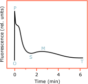
If a photosynthetic sample is kept for some time in the dark and then illuminated, its fluorescence will first rise quickly with time, then will slowly diminish and after one or several transient peaks will eventually reach a steady-state. This phenomeon is called fluorescence induction and was first discovered by Kautsky and Hirsch (1931). The graphical representation of the fluorescence intensity versus time is the fluorescence induction curve (Fig. 5). The shape and the amplitude of this curve tell a lot about the processes that take place during the induction period and about the physiological state of the sample.
There are many excellent books and reviews about fluorescence induction. The reader might refer to the reviews by Krause and Weis (1991), Govindjee (1995), Lazár (1999), and Strasser et al. (2000). Here, this topic will be treated very briefly.
In the dark-adapted plant samples the electron carriers of the electron transport chain are oxidised and the PS2 reaction centres are in their open state. Upon illumination, the open reaction centres conduct photochemical reactions with high efficiency. That is why the initial level of fluorescence Fo (corresponding to point O of the FIC) is low. The product of the photochemical reactions are electrons accumulated on the electron carriers — QA, QB, etc. With the reduction of the electron acceptors, the PS2 reaction centres are closed and fluorescence rises. This is the O-P section of the FIC and the fluorescence emitted by the closed reaction centres is called variable fluorescence. If the intensity of the actinic light is high enough (saturating), the point will be reached when all reaction centres are closed (all QA are reduced). The reason for this is that the remaining part of the photosynthetic chain — PS1 and the dark reactions — is inactive in the dark-adapted state and needs some time to activate.
With activation of PS1 and the dark phase of photosynthesis, the electrons stored in the pool of acceptors are withdrawn and the PS2 reaction centres are reopened. As a consequence the fluorescence decreases as the P-S-M-T part of the FIC shows. This is called photochemical quenching, because the fluorescence decreases at the expense of increased photochemistry. If the actinic light intensity is higher, so that the photosynthetic chain cannot keep up with it, a number of protective mechanisms come into play which dissipate the excessive light energy as heat. These processes also decrease the fluorescence intensity and are termed non-photochemical quenching. In reality, the drop of fluorescence after the peak P is due to both photochemical and non-photochemical quenching, with the latter playing a major role at high light conditions.
Knowing the mechanisms that form each phase of the induction transient, we can draw information from this transient about the underlying photosynthetic reactions. For example, the relative amplitude of the variable fluorescence (P−O)/O is a measure of the yield of the photochemical reaction; the area below the fluorescence curve is proportional to the number of reduced electron acceptors, and the complementary area (above the curve) is a measure of the size of the available pool of acceptors; the amplitude of the fluorescence decrease (quenching) can tell about the dark reactions of photosynthesis and so on.
